Microtubules displaying centrosomal and noncentrosomal pattern play vital roles in cell migration, cell division, vesicle transport and many other essential biological processes. Noncentrosomal microtubules exist in versatile cell types including epithelial cells, neurons, and myocytes. In mammalian epithelial cells, the CAMSAP family of proteins has been identified as a group of regulators of noncentrosomal microtubule minus ends. Previously scientists have proved that CAMSAP3, a member of the CAMSAP family, localizes at the noncentrosomal microtubule minus end and stabilizes microtubule, however, little is known about the mechanism and function for the anchor of noncentrosomal microtubule minus end.
In a recent study, researchers from MENG Wenxiang’s group at the Institute of Genetics and Developmental Biology, Chinese Academy of Sciences, first uncovered the mechanism for the noncentrosomal microtubule minus end anchoring at F-actin via a CAMSAP3-related complex with new structure foundation of actin and microtubule.
They found in the beginning that in Caco2 epithelial cells, CAMSAP3 stays quite stable at an unknown position in the cell depending on its CC(1, 2) domain. Upon mass spectrometric analysis and immunoprecipitation they proved that CAMSAP3 interacts with the 19th spectrin domain of ACF7 (actin-microtubule crosslinking family 7) through its CC (1, 2) domain. ACF7 is reported as a microtubule plus end protein that can interact with EB1, a protein that binds the microtubule plus ends, while surprisingly they found that ACF7 localized at the minus end of noncentrosomal microtubules depending on CAMSAP3.
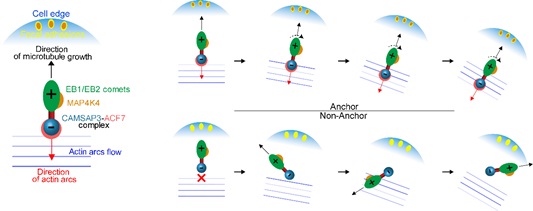
Furthermore, they proved that ACF7 anchors noncentrosomal microtubule minus end at F-actin through two CH domains at its N-terminal. In the leading edge of migrating cells, Myosin II facilitates stress fiber formation and F-actin undergoes retrograde flow, in this process, they detected that noncentrosomal microtubule minus end CAMSAP3 anchoring at F-actin forces it flowing together with actin arcs into the cell center, and its flow depends on ACF7 and Myosin II activity. This uncovered the reason for the coordinated retrograde flow of microtubule and F-actin at the cell leading edge.
Coordination of the retrograde flow of CAMSAP3 and F-actin is essential for the regulation of CAMSAP3 localization and maintaining proper length of parallel microtubules at the leading edge. And the failed anchor of noncentrosomal microtubules at F-actin led to inconsistent orientation of EB2 emerged from CAMSAP3 sites, which resulted in decreased targeting frequency of EB2 to focal adhesions, and perturbed both MAP4K4 location at focal adhesions and cell migration.
Taken together, they first uncovered the mechanism for the noncentrosomal microtubule minus end anchoring at F-actin via CAMSAP3-ACF7 complex which is the new structure foundation of actin and microtubule, and found ACF7 locates at the minus end of noncentrosomal microtubules depending on CAMSAP3. They also described the reason for the coordination of noncentrosomal microtubules retrograde flow with actin arcs, and finally verified the essential role for the anchor of noncentrosomal microtubules in cell migration.
All of these conclusions provide new insights for further functional research of noncentrosomal microtubules in epithelia and neurons.
This work, entitled “the CAMSAP3-ACF7 Complex Couples Noncentrosomal Microtubules with Actin Filaments to Coordinate Their Dynamics”, has been published on
Developmental Cell as the cover paper (
http://dx.doi.org/10.1016/j.devcel.2016.09.003).
This work was supported by the National Natural Science Foundation of China, the National Basic Research Program of China, and the Key Research Program of the Chinese Academy of Sciences.
Noncentrosomal microtubules minus-ends anchor at actin filaments via the CAMSAP3-ACF7 complex and contribute to cell migration (Image by IGDB)
10 October 2016
Volume 39, Issue 1
On the cover: An artist's depiction of the Chinese mythological figure Nezha holding a red armillary sash (representing microtubules) and riding on wind fire wheels (representing ACF7) over ocean waves (representing actin flow) to conquer the Dragon King. To learn more about how Nezha/CAMPSAP3 at the minus ends of non-centrosomal microtubules works with ACF7 to anchor microtubules to actin filaments and how these microtubules orient using retrograde actin flow to facilitate cell migration, see Ning, Yu, et al., pp. 61–74. (Image by SHEN Guangwei).
Contact:
Dr. MENG Wenxiang
 Noncentrosomal microtubules minus-ends anchor at actin filaments via the CAMSAP3-ACF7 complex and contribute to cell migration (Image by IGDB)
 10 October 2016Volume 39, Issue 1On the cover: An artist's depiction of the Chinese mythological figure Nezha holding a red armillary sash (representing microtubules) and riding on wind fire wheels (representing ACF7) over ocean waves (representing actin flow) to conquer the Dragon King. To learn more about how Nezha/CAMPSAP3 at the minus ends of non-centrosomal microtubules works with ACF7 to anchor microtubules to actin filaments and how these microtubules orient using retrograde actin flow to facilitate cell migration, see Ning, Yu, et al., pp. 61–74. (Image by SHEN Guangwei).Contact:Dr. MENG WenxiangEmail: wxmeng@genetics.ac.cn
 CAS
CAS
 中文
中文


.png)
