Lipids are structural components of cellular membranes and signaling molecules that are widely involved in development and disease, the glycosphingolipids (GSLs) are particularly abundant in the nervous system and are essential for brain development. However the underlying molecular mechanisms are poorly understood, partly because of the vast variety of lipid species and complexity of synthetic and turnover pathways. There is usually no linear relationship between genes and lipid variants, making it difficult to specifically dissect the function of a specific lipid by genetic means.
A team led by Dr. ZHANG Yong Q. from the Institute of Genetics and Developmental Biology of the Chinese Academy of Sciences, taking advantages of effective genetic manipulations and less redundant metabolic pathways in Drosophila, investigated the role of individual lipids in synaptic development by a genetic screen.
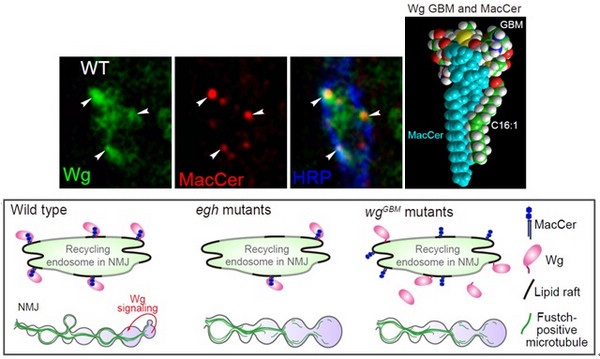
They identifies that mannosyl glucosylceramide (MacCer), a species GSL, promotes synaptic bouton formation at the neuromuscular synapse. MacCer co-localizes with growth factor Wnt1/Wingless (Wg) and positively regulates its synaptic level and facilitates presynaptic Wg signaling.
Furthermore, in cooperation with Professor Jacques Fantini from Aix-Marseille University, they found a functional GSL-binding motif in Wg exhibiting a high affinity for MacCer by molecular simulation and in vitro biochemistry. Further in vivo studies revealed that this motif is required for MacCer-Wg co-localization and normal synaptic growth.
This is the first report defining a specific sphingolipid in controlling synapse growth and revealing a novel mechanism whereby the MacCer promotes synaptic bouton formation via Wg signaling.
This paper entitled “The glycosphingolipid MacCer promotes synaptic bouton formation in
Drosophila by interacting with Wnt” has been published in
eLife on Oct. 25
TH (DOI:
https://doi.org/10.7554/eLife.38183.001).
This work was supported by Ministry of Science and Technology of China, National Natural Science Foundation of China, and the Chinese Academy of Sciences.
Immunostaining image showing the co-localization between MacCer and Wg (upper left); molecular simulation showing a MacCer-Wg binding (upper right); a schematic figure illustrates that MacCer regulates Wg signaling during synapse growth (lower panel). (Image by IGDB)
Contact:
Lei Qi
Institute of Genetics and Developmental Biology , Chinese Academy of Sciences
E-mail: lqi@genetics.ac.cn
 Immunostaining image showing the co-localization between MacCer and Wg (upper left); molecular simulation showing a MacCer-Wg binding (upper right); a schematic figure illustrates that MacCer regulates Wg signaling during synapse growth (lower panel). (Image by IGDB)Contact:Lei QiInstitute of Genetics and Developmental Biology , Chinese Academy of SciencesE-mail: lqi@genetics.ac.cn
 CAS
CAS
 中文
中文


.png)
