Using liverwort (Marchantia polymorpha) as a model, researchers led by Prof. LI Hongju from the Institute of Genetics and Developmental Biology of the Chinese Academy of Sciences have explored the molecular mechanism of sperm release in bryophytes.
The study was published in Nature Plants (DOI: 10.1038/s41477-024-01703-1).
Sexual reproduction is essential for the environmental adaptation and evolution of plants. Unlike angiosperms, which rely on pollen tube growth to deliver immotile sperm cells to the embryo sac for fertilization, motile sperm in the basal land plants, bryophytes, are released into the water and swim to the egg cell in the archegonia. While the fertilization process in bryophytes has intrigued many researchers, the factors that regulate sperm release have remained unknown.
In this study, the researchers used RNA sequencing and analysis to identify four Mildew Resistance Locus O (MpMLO) proteins that are specifically expressed in the reproductive tissues of Marchantia. Expression pattern analysis revealed that these MpMLOs are expressed only in antheridia, which contain the sperm cells.
Using the CRISPR/Cas9 system, the researchers generated the Mpmlo1 mutant, which failed to release sperm. Subcellular localization revealed that MpMLO1-Citrine first localizes to the plasma membrane of tip cells at the end of the antheridia jacket layer, which causes cell death of these cells.
In contrast, the researchers found that tip cells in the Mpmlo1 mutant do not undergo cell death after antheridia maturation and continue to enlarge even after antheridia degeneration due to antheridia aging. By introducing the Ca2+ sensor R-GECO1 into both wild-type and Mpmlo1 mutant plants, the researchers were able to study the dynamic variation of Ca2+ in antheridia jacket cells.
They recorded high Ca2+ levels in the tip cells that burst open to release the sperm, while they found reduced cytoplasmic Ca2+ levels in the tip cells of the Mpmlo1 mutant that failed to release sperm.
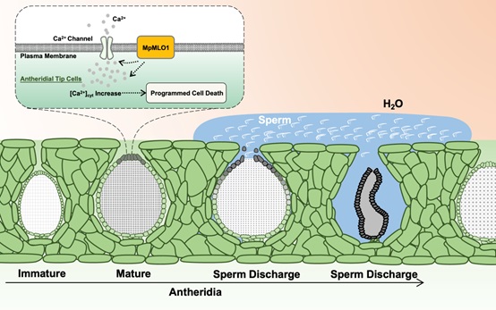
In conclusion, the researchers found that programmed cell death (PCD) is a prerequisite for sperm release from Marchantia antheridia. MpMLO1, expressed in tip cells, increases the cytoplasmic Ca2+ levels and induces PCD in these cells. Subsequent water entry into the antheridial pore transports the sperm mucilage to the receptacle surface for further fertilization.
This study sheds light on the molecular basis of sperm discharge in ancestral land plants and highlights the evolutionary conservation of the MLO-Ca2+ signaling module, which can be traced back to the last common ancestor of liverworts and flowering plants.
Figure. MpMLO1 localized on the plasma membrane of tip cells induces cytoplasmic Ca2+ increase, triggering PCD in these cells. (Image by LI Hongju's group)
Contact:
Dr. LI Hongju
Institute of Genetics and Developmental Biology, Chinese Academy of Sciences
Email: hjli@genetics.ac.cn
 Figure. MpMLO1 localized on the plasma membrane of tip cells induces cytoplasmic Ca2+ increase, triggering PCD in these cells. (Image by LI Hongju's group)Contact:Dr. LI HongjuInstitute of Genetics and Developmental Biology, Chinese Academy of SciencesEmail: hjli@genetics.ac.cn
 CAS
CAS
 中文
中文


.png)
