On July 30, 2025, the research team led by YANG Weicai at the Institute of Genetics and Developmental Biology, Chinese Academy of Sciences, using the model plant Arabidopsis thaliana, elucidated how plant cells regulate membrane curvature and morphological remodeling through lipid bilayer asymmetry and the interleaflet coupling of different phospholipid species. They further uncovered the molecular mechanism by which phosphatidylinositides drive cell plate structural transitions during cytokinesis, ensuring successful daughter cell separation.
The study showed that knockout of myo-inositol-1-phosphate synthase (MIPS), which reduces phosphatidylinositide levels, led to significantly shortened roots, increased polyploidy, and incomplete cytokinesis. Transmission electron microscopy and spinning-disk confocal microscopy revealed that mips mutant had discontinuous, thickened cell plates and prolonged division time. Structural instability during lateral expansion caused cell plate holes and incomplete separation.
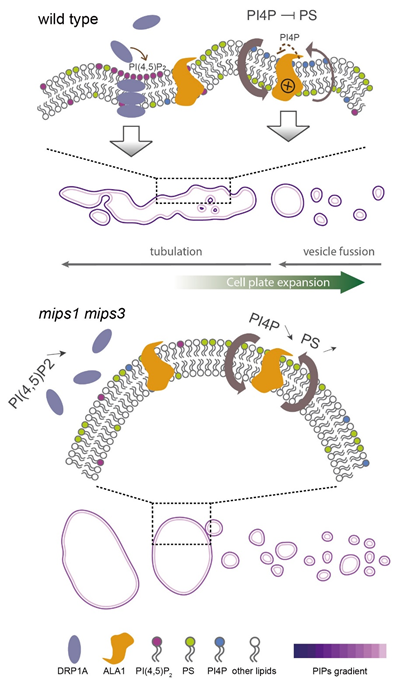
Through EMS mutagenesis and suppressor screening, the researchers found that Aminophospholipid ATPase 1 (ALA1) knockout could restore cytokinesis in mips mutant. Fluorescent lipid labeling confirmed that ALA1 flips phosphatidylserine (PS), and treatment with the phosphatidylinositol-4-phosphate (PI4P) synthesis inhibitor phenylarsine oxide (PAO) demonstrated that PI4Pinhibits this flipping activity. Super-resolution confocal microscopy showed discontinuous PS distribution in wild type cell plates but continuous and elevated PS levels in mips mutant, suggesting that PI4P prevents excessive PS accumulation on the cytoplasmic leaflet.
Because flippases transport lipids from the outer to the inner leaflet, excessive flipping increases cytoplasmic membrane curvature. Both MIPS mutation and PAO treatment elevated curvature at the cell plate, causing thickening and protrusions. Importantly, this effect was not limited to the cell plate—overall plasma membrane curvature on the cytoplasmic side was also significantly elevated, forming invaginations and large endocytic vesicles. Using bio-layer interferometry (BLI), the researchers confirmed that ALA1 binds PI4P, supporting a model in which PI4P inhibits ALA1 to reduce local curvature and promote cell plate flattening.
Unexpectedly, overexpression of DRP1A, introduced as a fluorescent marker, partially rescued the mips cytokinesis defect. DRP1A, a dynamin-family GTPase, remodels membranes via GTP hydrolysis and participates in endocytosis, vesicle budding, organelle fission, and cytokinesis. Super-resolution confocal imaging showed that DRP1A formed discrete foci and contractile ring-like structures in wild type cells, but these foci were fewer in number and the rings appeared enlarged in the mutant background. Results from BLI and PI(4,5)P₂ depletion experiments revealed that PI(4,5)P₂ binds to DRP1A and promotes membrane constriction, aiding cell plate maturation.
In conclusion, PI4P reduces membrane curvature by inhibiting ALA1-mediated PS flipping, while PI(4,5)P₂ recruits DRP1A to drive constriction. Together, these phosphoinositides coordinate membrane remodeling to ensure successful plant cytokinesis.
Contact:
Dr. LUO Yu
Email: luoyu@genetics.ac.cn
Prof. YANG Weicai
Email: wcyang@genetics.ac.cn
Institute of Genetics and Developmental Biology, Chinese Academy of Sciences
 CAS
CAS
 中文
中文


.png)