DNA is not randomly distributed within the nucleus but is precisely folded into complex three-dimensional structures. Our understanding of the dynamic regulation of higher-order chromatin architecture remains limited. For instance, condensed chromatin tends to fragment during tumorigenesis, whereas it tends to fuse during neural aging. Research on the dynamic behavior of higher-order chromatin architecture is still in its infancy.
In recent years, single-cell 3D genomics technologies have greatly advanced our understanding of chromatin conformation, yet these methods cannot preserve the spatial position of cells within tissues, making it difficult to study how genome architecture regulates gene expression in native cellular environments. Recently developed spatial 3D genomics technologies have adopted ligation-based Hi-C strategies, which however preferentially detect short-range chromatin interactions (within 10 Mb).
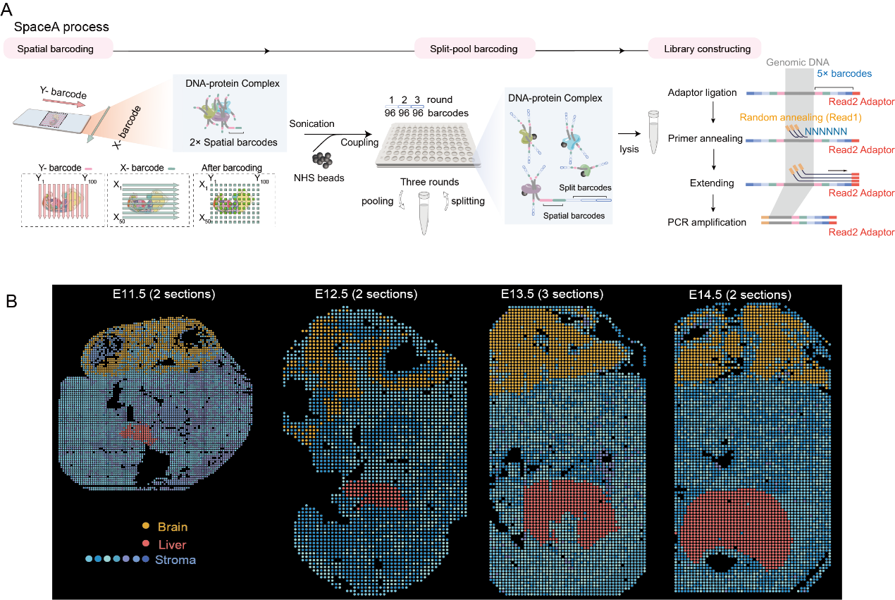
Now, a group led by Prof. LI Xiang from the Institute of Genetics and Developmental Biology (IGDB), Chinese Academy of Sciences, developed SpaceA, a spatial 3D genomics technology that enables high-confidence, in situ profiling of long-range chromatin interactions within tissue sections, through integrating microfluidic spatial barcoding with a SPRITE-like ligation-free contact detection strategy.
The researchers applied SpaceA to nine mouse embryo sections spanning four developmental stages (E11.5 to E14.5), generating approximately 4 TB of deeply sequenced data and systematically resolving the dynamic changes in chromatin conformation during embryonic development.
One key finding is that A/B compartments undergo a dramatic transition at E12.5–E13.5, coinciding with profound transcriptomic changes. Another key finding is the discovery, for the first time, of a unique chromatin assembly mechanism in the embryonic liver that operates independently of classical A/B compartment determination: mid-range interactions (10–40 Mb) are significantly enhanced, interactions between adjacent compartments are strengthened, while long-range inter-chromosomal interactions (>40 Mb) are weakened. Microscopy imaging revealed heterochromatin adhesion in liver cell nuclei, confirming this unique chromatin structure, which is accompanied by a global decline in gene expression. Notably, this differs from the classical model of promoting gene expression through A/B compartment switching or intra-compartment interactions.
This study provides a powerful new tool for studying higher-order chromatin architecture in native tissue environments and facilitates the discovery of novel mechanisms governing higher-order chromatin structure.
SpaceA resolves the dynamic changes in chromatin conformation during mouse embryonic development (Image by IGDB). A. Schematic workflow of SpaceA. B. Spatial distribution of mouse embryo sections clustered based on chromatin interaction data.
Contact:
Prof. LI Xiang
Institute of Genetics and Developmental Biology, Chinese Academy of Sciences
Email: lixiang@genetics.ac.cn
 CAS
CAS
 中文
中文



.png)
 SpaceA resolves the dynamic changes in chromatin conformation during mouse embryonic development (Image by IGDB). A. Schematic workflow of SpaceA. B. Spatial distribution of mouse embryo sections clustered based on chromatin interaction data.Contact:Prof. LI XiangInstitute of Genetics and Developmental Biology, Chinese Academy of SciencesEmail: lixiang@genetics.ac.cn
