Selected Publications
# Co-first author
* Corresponding author
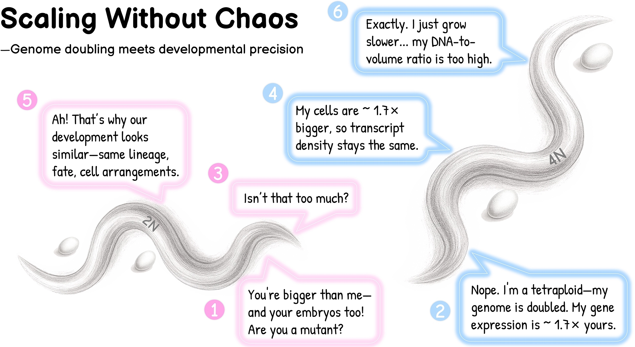
Intracellular buffering enables developmental robustness after genome doubling in C. elegans embryos. Yang M*, Bai Y, Wang Z, Du Z*. Cell Reports. 2026 Mar 2:117005.
Previewed in Cell Reports by Alexander Lessenger and Rebecca Heald: "Building life from a bigger blueprint: Embryogenesis in whole-organism tetraploids."
To see and to know: the power of live imaging in illuminating and decoding biological complexity. Yang M*, Du Z*. Journal of Genetics and Genomics. 2026 Mar;53(3):361-380.
Reaffirming the value of model organisms in training scientific minds. Yang M, Du Z*. Nature Cell Biology. 2025 Oct;27(10):1589-1591.
A journey into biological complexity: continuing the legacy of Doug and Bill. Yang M, Du Z*.Hereditas (Beijing). 2025 Dec;47(12):1377-1386.
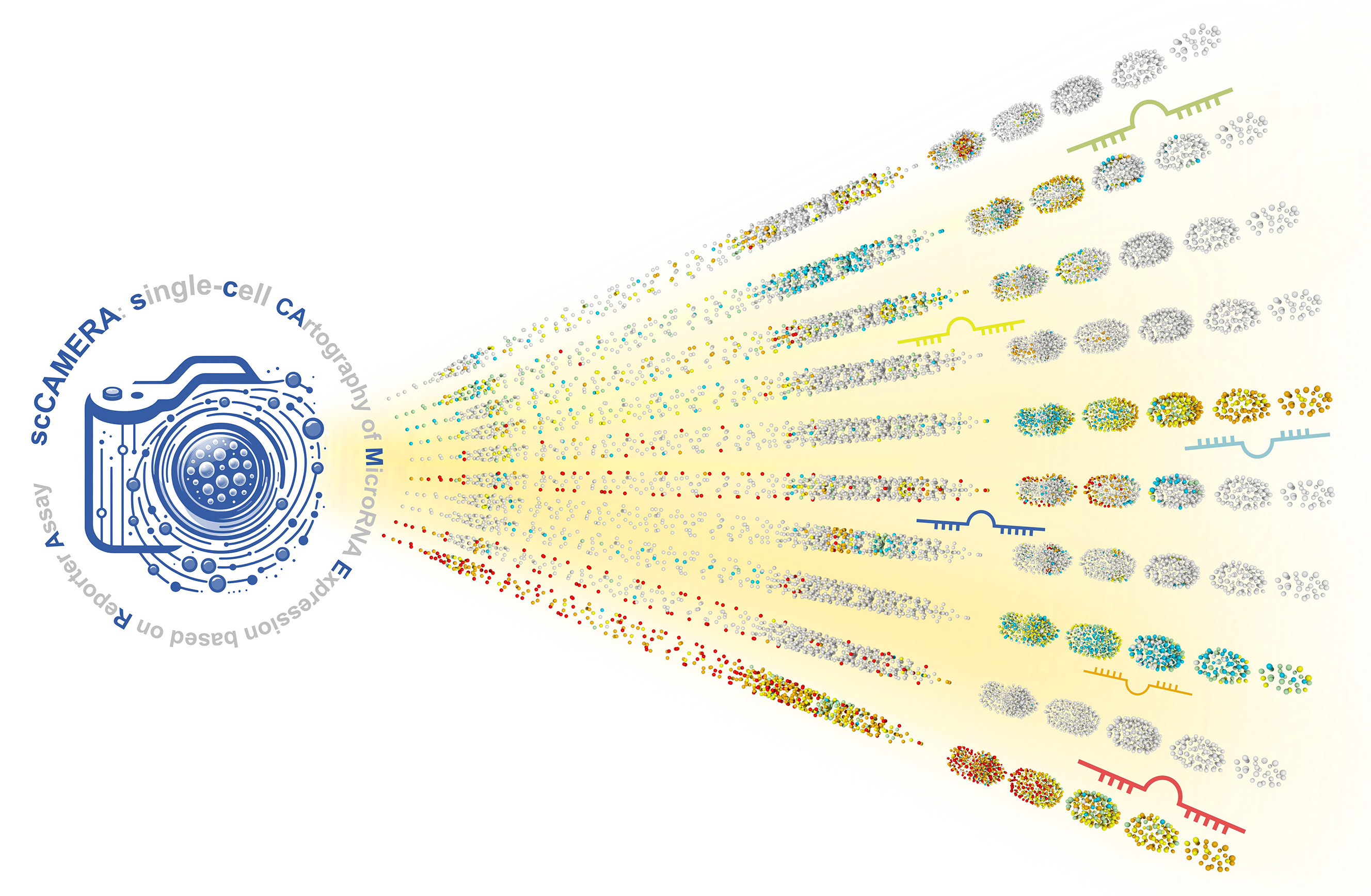
A lineage-resolved cartography of microRNA promoter activity in C. elegans empowers multidimensional developmental analysis. Xu W#, Liu J#, Qi H#, Si R, Zhao Z, Tao Z, Bai Y, Hu S, Sun X, Cong Y, Zhang H, Fan D, Xiao L, Wang Y, Li Y*, Du Z*. Nature Communications. 2024 Mar 30;15(1):2783.
Spatiotemporal analysis of mRNA-protein relationships enhances transcriptome-based developmental inference. Fan D, Cong Y, Liu J, Zhang H, Du Z*. Cell Reports. 2024 Mar 26;43(3):113928 (1-23).
Defect-buffering cellular plasticity increases robustness of metazoan embryogenesis. Xiao L, Fan D, Qi H, Cong Y, Du Z*. Cell Systems. 2022 Aug 17;13(8):615-630.
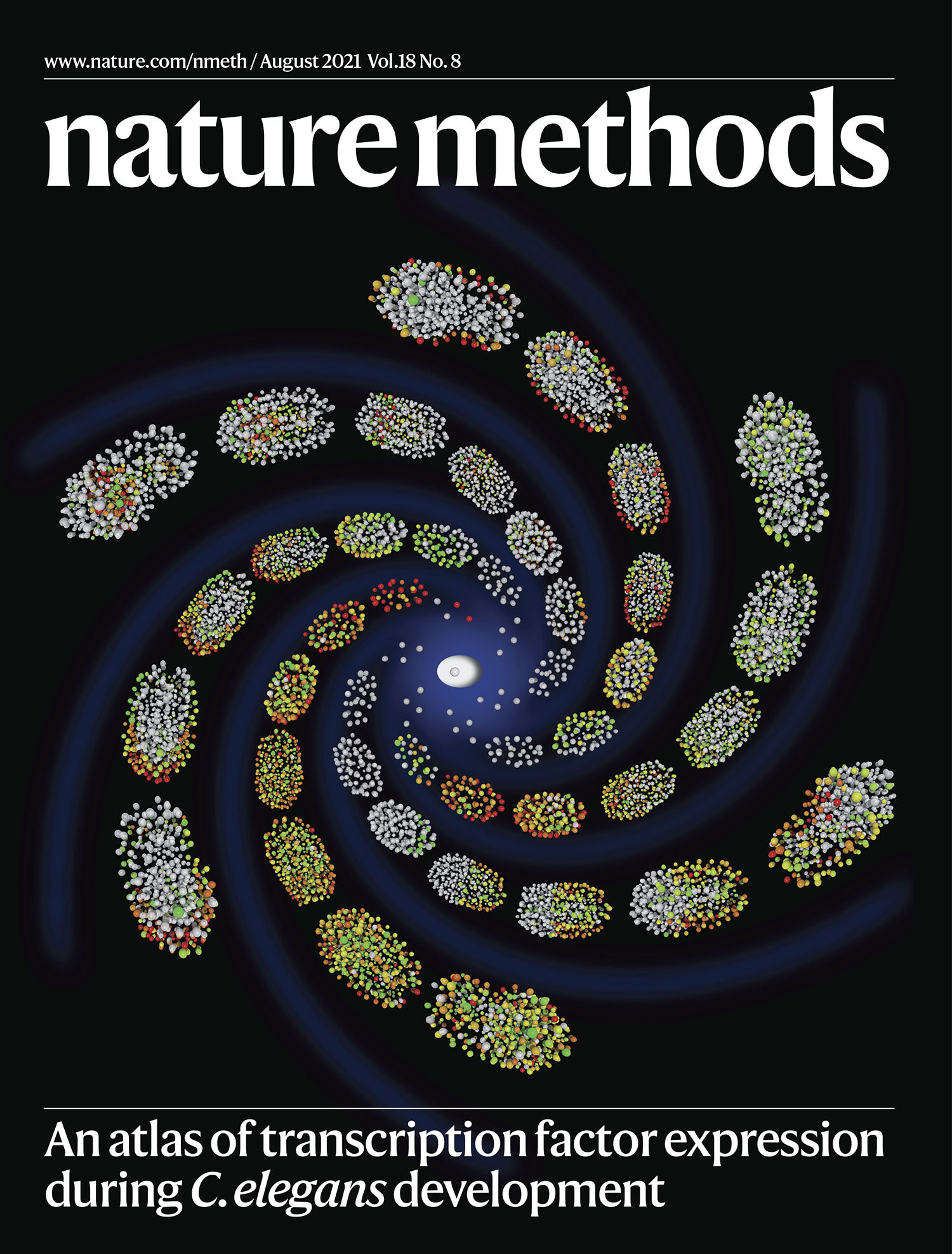
A 4D single-cell protein at las of transcription factors delineates spatiotemporal patterning during embryogenesis. Ma X#, Zhao Z#, Xiao L#, Xu W, Kou Y, Zhang Y, Wu G, Wang Y, Du Z*. Nature Methods. 2021 Aug;18(8):893-902.
Cover story
Highlighted in a Nature Methods News & Views by Aly Makhlouf and Marta Shahbazi: "The long and winding road of development: a coordinated song of transcription factors."
Single-cell dynamics of chromatin activity during cell lineage differentiation in Caenorhabditis elegans embryos. Zhao Z#, Fan R#, Xu W, Kou Y, Wang Y, Ma X, Du Z*. Molecular Systems Biology. 2021 Apr;17(4):e10075 (1-24).
Systems properties and spatiotemporal regulation of cell position variability during embryogenesis. Li X#, Zhao Z, Xu W, Fan R, Xiao L, Ma X, Du Z*. Cell Reports. 2019 Jan 8;26(2):313-321.e7.
Digital development: a database of cell lineage differentiation in C. elegans with lineage phenotypes, cell-specific gene functions and a multiscale model. Santella A, Kovacevic I, Herndon LA, Hall DH, Du Z*, Bao Z*. Nucleic Acids Research. 2016 Jan 4;44(D1):D781-5.
The regulatory landscape of lineage differentiation in a metazoan embryo. Du Z*, Santella A, He F, Shah PK, Kamikawa Y, Bao Z*. Developmental Cell. 2015 Sep 14;34(5):592-607.
De novo inference of systems-level mechanistic models of development from live-imaging-based phenotype analysis. Du Z, Santella A, He F, Tiongson M, Bao Z*. Cell. 2014 Jan 16;156(1-2):359-72.
Other Publications
Lactate shuttling links histone lactylation to adult hippocampal neurogenesis in mice. Li Z, Liang Z, Qi H, Luo X, Wang M, Du Z, Guo W*. Developmental Cell. 2025 Apr 21;60(8):1182-1198.
Vacuolar H+-ATPase determines daughter cell fates through asymmetric segregation of the nucleosome remodeling and deacetylase complex. Xie Z, Chai Y*, Zhu Z, Shen Z, Guo Z, Zhao Z, Xiao L, Du Z, Ou G, Li W*. Elife. 2024 Jul 12;12:RP89032.
Gene reglatory patterning codes in early cell fate specification of the C. elegans embryo. Cole AG#, Hashimshony T#, Du Z, Yanai I*. Elife. 2024 Jan 29;12:RP87099.
Metabolic plasticity sustains the robustness of Caenorhabditis elegans embryogenesis. Chen S, Su X, Zhu J, Xiao L, Cong Y, Yang L, Du Z, Huang X*. EMBO Reports. 2023 Dec 6;24(12):e57440.
Dynamic chromatin regulatory programs during embryogenesis of hexaploid wheat. Zhao L, Yang Y, Chen J, Lin X, Zhang H, Wang H, Wang H, Bie X, Jiang J, Feng X, Fu X, Zhang X, Du Z, Xiao J*. Genome Biology. 2023 Jan 13;24(1):7.
Comparative proteome and cis-regulatory element analysis reveals specific molecular pathways conserved in dog and human brains. Hong H#, Zhao Z#, Huang X, Guo C, Zhao H, Wang GD, Zhang YP, Zhao JP, Shi J, Wu QF, Jiang YH, Wang Y, Li LM, Du Z, Zhang YQ*, Xiong Y*. Mol Cell Proteomics. 2022 Aug;21(8):100261.
Dogs lacking Apolipoprotein E show advanced atherosclerosis leading to apparent clinical complications. Zhao H#, Zhao J#, Wu D#, Sun Z#, Hua Y, Zheng M, Liu Y, Yang Q, Huang X, Li Y, Piao Y, Wang Y, Lam SM, Xu H, Shui G, Wang Y, Yao H, Lai L, Du Z, Mi J*, Liu E*, Ji X*, Zhang YQ*. Science China Life Science. 2022 Jul;65(7):1342-1356.
Multivariable regulation of gene expression plasticity in metazoans. Xiao L#, Zhao Z#, He F*, Du Z*. Open Biology. 2019 Dec;9(12):190150 (1-16).
Lineage context switches the function of a C. elegans Pax6 homolog in determining a neuronal fate. Brandt JP#, Rossillo M#, Du Z, Ichikawa D, Barnes K, Chen A, Noyes M, Bao Z, Ringstad N*. Development. 2019 Apr 15;146(8):dev168153.
mTOR regulates phase separation of PGL granules to modulate their autophagic degradation. Zhang G, Wang Z, Du Z, Zhang H*. Cell. 2018 Sep 6;174(6):1492-1506.e22.
Trans-splicing enhances translational efficiency in C. elegans. Yang YF#, Zhang X#, Ma X#, Zhao T#, Sun Q, Huan Q, Wu S, Du Z*, Qian W*. Genome Research. 2017 Sep;27(9):1525-1535.
E3 ubiquitin ligases promote progression of differentiation during C. elegans embryogenesis. Du Z#, He F#, Yu Z, Bowerman B, Bao Z*. Developmental Biology. 2015 Feb 15;398(2):267-79.
POS-1 promotes endo-mesoderm development by inhibiting the cytoplasmic polyadenylation of neg-1 mRNA. Elewa A, Shirayama M, Kaymak E, Harrison PF, Powell DR, Du Z, Chute CD, Woolf H, Yi D, Ishidate T, Srinivasan J, Bao Z, Beilharz TH, Ryder SP, Mello CC*. Developmental Cell. 2015 Jul 6;34(1):108-18.
A semi-local neighborhood-based framework for probabilistic cell lineage tracing. Santella A, Du Z, Bao Z*. BMC Bioinformatics. 2014 Jun 25;15:217.
Systematic quantification of developmental phenotypes at single-cell resolution during embryogenesis. Moore JL, Du Z, Bao Z*. Development. 2013 Aug;140(15):3266-74.
Inverted selective plane illumination microscopy (iSPIM) enables coupled cell identity lineaging and neurodevelopmental imaging in Caenorhabditis elegans. Wu Y*, Ghitani A, Christensen R, Santella A, Du Z, Rondeau G, Bao Z, Colón-Ramos D, Shroff H. Proc Natl Acad Sci U S A. 2011 Oct 25;108(43):17708-13.
A hybrid blob-slice model for accurate and efficient detection of fluorescence labeled nuclei in 3D. Santella A, Du Z, Nowotschin S, Hadjantonakis AK, Bao Z*. BMC Bioinformatics. 2010 Nov 29;11:580.
Single-molecule analysis reveals changes in the DNA replication program for the POU5F1 locus upon human embryonic stem cell differentiation. Schultz SS, Desbordes SC, Du Z, Kosiyatrakul S, Lipchina I, Studer L, Schildkraut CL*. Molecular and Cellular Biology. 2010 Sep;30(18):4521-34.
Genome-wide colonization of gene regulatory elements by G4 DNA motifs. Du Z#, Zhao Y#, Li N*. Nucleic Acids Research. 2009 Nov;37(20):6784-98.
Genome-wide analysis reveals regulatory role of G4 DNA in gene transcription. Du Z#, Zhao Y#, Li N*. Genome Research. 2008 Feb;18(2):233-41.
Extensive selection for the enrichment of G4 DNA motifs in transcriptional regulatory regions of warm blooded animals. Zhao Y#, Du Z#, Li N*. FEBS Letters. 2007 May 15;581(10):1951-6.
Data/Resource Access and Visualization
C. elegans strains donated to the Caenorhabditis Genetics Center (CGC)
single-cell CArtography of MicroRNA Expression based on Reporter Assay (scCAMERA) (Nature Communications 2024)
Single-cell Phenotypic Landscape of Embryonic Development in Conserved Genes (Cell Systems 2022)
4D Single-Cell Protein Atlas of Transcription Factors (Nature Methods 2021)
Single-cell chromatin activity landscape (Molecular Systems Biology 2021)
Cell position variability during early embryogenesis (Cell Reports 2019)
Single-cell phenomics of cell lineage differentiation following perturbing essential genes (Cell 2014 and Developmental Cell 2015)
 CAS
CAS
 中文
中文


.png)