Sexual reproduction is the predominant reproduction style in nature. The most pivotal event of reproductive development in all species employing sexual reproduction is the transition from mitotic to meiotic cell cycle. However, the mechanisms orchestrating meiosis initiation remain elusive, particularly in plants. Angiosperms are heterosporous plants in which two kinds of spore mother cells are generated, with male and female male reproductive organs acquiring different developmental courses in spore genesis.
Scientists in Dr. Zhukuan Cheng’s group (from the Institute of Genetics and Developmental Biology, Chinese Academy of Sciences) show that plant pollen mother cells contain a specific meiosis initiation machinery through characterization of a rice gene MICROSPORELESS1 (MIL1). The mil1 mutant does not produce microspores in anthers but has the normal female fertility. Its anthers are defective in the meiotic entry of sporogenous cell progeniesand in the differentiation of surrounding somatic cell layers, resulting in locules filled with somatic cells instead of microspores. Thus, they suggest meiotic entry in microsporocytes is directed by an anther-specific mechanism, which requires MIL1 activity, and redox regulation might play important roles in this process.
This work with Lilan Hong, Ding Tang and Keming Zhu as the co-first authors has been online published on The Plant Cell (doi:10.1105/tpc.111.093740). This research was supported by grants from the Ministry of Science and Technology, National Natural Science Foundation of China.
AUTHOR CONTACT:
Zhukuan Cheng, Ph.D.
Institute of Genetics and Developmetnal Biology, Chinese Academy of Sciences, Beijing, China.
E-mail: zkcheng@genetics.ac.cn
(Image by Lilan Hong et al)
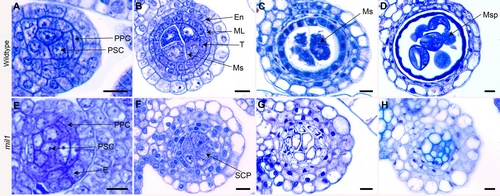
Figure Toluidine Blue-Stained Transverse Sections of Anthers in the Wild Type and mil1. (A) Wild type anthers with primary parietal cells and primary sporogenous cells. (B) Wild type anthers with microsporocytes and four parietal layers. (C) Wild type anthers at meiotic stage exhibiting crushed middle layer. (D) Wild type anthers filled with microspores, with middle layer and tapetum degraded. (E) mil1 anthers with primary parietal cells and primary sporogenous cells. (F) mil1 anthers with SCPs in the center. They generally have four parietal layers, but show extra layers in some regions. (G) mil1 SCPs increase in number and decrease in size as anthers develop. No cell degeneration is observed in wall cells. (H) mil1 anthers are composed of somatic cells. Scale bars, 10 μm.