The brain devotes a substantial portion of its net energy expenditure into maintaining membrane lipid dynamics, which essentially entails the perseverance of a unique fatty acid profile esterified across various classes of membrane lipids, including phospholipids and sphingolipids. A precise distribution of fatty acyl heterogeneity across various membrane lipid classes would, therefore, be of immense importance in maintaining membrane lipid integrity and proper functioning of the brain. On basis of their high genetic homology to humans (c.a. 92.5% to 95%), the Rhesus macaques share a remarkably similar profile of normative aging in terms of age-related phenotypes. Despite the considerable research efforts dedicated to unraveling the neurological impacts of polyunsaturated fatty acids (PUFAs) to human brain aging; and that several clinical studies have clearly established a changing pattern of docosahexaenoic acids (DHAs) homeostasis during normative aging in humans, no clear counterpart has been observed in animal or in vitro studies. The lack of a suitable animal model, coupled with the ethical constraints in working with human brain tissues, has largely circumscribed the arena of PUFA brain research.
In a recent study, using state-of-the-art lipidomic approach, SHUI Guanghou’s group from the Institute of Genetics and Developmental Biology, Chinese Academy of Sciences, reported herein an extensive lipidomic atlas of the changing membrane lipid landscape in the frontal cortex of Rhesus macaques across three selected age groups (i.e. young, sexually-mature and old) to the details of individual fatty acyls. In particular, the use of electrospray ionization in their approach has enabled the detection of phospholipids, sphingolipids and glycerolipids in their intact forms, thereby preserving the head group information pertaining to the temporal changes in individual fatty acyl with aging. The fatty acyl-specific analysis of the monkey frontal cortical membrane lipidome strongly justified the suitability of Rhesus macaques as a model for studying normative brain aging in humans, especially with regard to brain PUFA metabolism, since age-dependent PUFA alterations previously reported in humans using gas chromatography-mass spectrometry (but with no details of head group information) are fully recapitulated in this animal model.
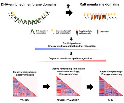
Remarkably, the lipidomic analysis revealed an intriguing pattern of PUFA-esterification across phospholipids on a temporal scale, with DHA displaying notable accretions in sexually-mature macaques for all phospholipid classes examined. This accumulation is distinct to DHA per se and not observable in all remaining PUFAs. On the other hand, arachidonic acid (ARA) exhibited sharp attritions in the membrane lipidomes of sexually-mature macaques, a decline which was attenuated only for cardiolipins (CLs). Interestingly, DHA enrichment in phospholipids was lost in old macaques, with accompanying augmentations in very-long-chain sphingomyelins (VLC-SMs). Correlation matrix analysis demonstrated an escalating degree of membrane lipid co-regulation with aging. In particular, strong co-regulation existed between CLs and PUFA-phospholipids, while DHA-phospholipids were negatively associated with the level of VLC-SMs in old macaques. The lipidomics data therefore point to an attractive possibility that a complex temporal interplay between DHA-enriched membrane microdomains and SM-/cholesterol-rich rafts may exist in neural membranes.
The lipid co-regulation analyses also suggest the temporal switching of functional membrane microdomains (DHA-enriched to SM-/cholesterol-rich raft membrane microdomains) may alter the efficiency of CL remodeling, leading to a gradual reduction in the level of total CLs and decline in energy availability from mitochondrial oxidative phosphorylation. The diminished energy supply, probably in concert with an altered membrane topology, may herald in alternative phospholipid synthetic pathways in place of de novo biosynthesis to maintainmembrane dynamics in a more energy-saving manner, which may explain the intense degree of membrane lipid co-regulation unique to old macaques.Central to this idea, it appears that the key event that initiates brain aging may lie essentially in a failure in maintaining a high level of esterified DHAs in neural membrane lipids. In addition, the loss of lipid co-regulation between LPC-22:6 and LPE-22:6 with phospholipids comprising DHAs in old macaques also indicated that the reductions in DHA-phospholipids may not be primarily attributed to a lack of DHA substrates supplied in the form of lysophospholipids across the blood brain barrier, but a failure to incorporate these DHA substrates into membrane lipids instead.
Interestingly, such mechanistic presumption is aligned with the clinical observations that while DHA diet supplementation is effective in preventing or delaying the onset of neurodegenerative diseases, no therapeutic effect of DHA supplementation was found for the treatment of established Alzheimer’s disease; which suggests that a breakdown in DHA esterification into neural membranes may prove more detrimental than a diminished dietary supply of DHA per se.
Figure 1: Schematic diagram illustrating the proposed role of changing membrane lipid dynamics that governs normative brain aging in Rhesus macaques. (Image by IGDB)
This study entitled “Biological relevance of fatty acyl heterogeneity to the neural membrane dynamics of Rhesus macaques during normative aging” was published online in Oncotarget (DOI: 10.18632/oncotarget.11190).
Contact:
Dr. SHUI Guanghou
Email: ghshui@genetics.ac.cn