Autism spectrum disorder (ASD) is a group of neurodevelopmental disorders characterized by impaired communication, reciprocal social interactions, and restricted and repetitive patterns of behaviors and interests. Human genetic studies show that mutations in SH3 domain and ankyrin repeat containing protein (SHANK) family members are causative for idiopathic ASD. SHANK is one of the most intensively studied ASD related protein at the postsynaptic density.
To study the function of SHANK, a group led by Prof. ZHANG Yongqing at the Institute of Genetics and Developmental Biology, Chinese Academy of Sciences, in collaboration with Dr. GAN Guangming at Southeast University, Professor LI Yan at Institute of Biophysics, Chinese Academy of Sciences, Professor JIN Shan at Hubei University, Professor HUANG Juan at Nanjing Medical University, Professor Ulrich Thomas at Leibniz Institute for Neurobiology, and Professor Yong-hui Jiang at Duke Medical School, generated Drosophila mutants of Shank, the only homolog of mammalian SHANK gene family.
They found that Shank expresses at the presynapse of the peripheral neuromuscular junction (NMJ), while Shank enriches in the neuropil and mushroom body calyx of the central nervous system. Specifically, Shank co-expresses with the postsynaptic density (PSD) protein Homer in the mushroom body calyces.
They observed structural defects in the mushroom body calyx of Shank mutants by anti-choline acetyltransferase (ChAT) staining, which labels presynaptic boutons of projection neuron (PN) termini in the calyx. Wild-type larva or adult brain showed evenly distributed ChAT positive puncta within glomeruli with multiple protrusions (Figure 1 left). The protrusions marked by ChAT positive puncta were lost and instead fused together to form smaller, uni-lobed glomeruli in Shank mutants (Figure 1 right).
To examine if Shank functions at pre- or post-synapse, they performed cell-type specific rescue experiment with pre- or post-synaptic specific Gal4 in the mushroom body calyces. Structural defects in mushroom body calyces of Shank mutants were not rescued by postsynaptic re-expression of Shank, but could be partially rescued by presynaptic re-expression of Shank; both pre and post re-expression generated stronger rescue effect compared with presynaptic re-expression of Shank alone. These results indicate a presynaptic function of Shank is required for the normal synaptic contacts at the mushroom body calyx. Electron microscopy analysis further showed ultra-structural defects in adult Shank mutants’ calyx; synaptic clefts were collapsed in Shank mutants, consistent with the light microscopy analysis results. They also examined Shank mutants’ behaviors; Drosophila Shank mutants showed reduced motor ability and defective olfactory acuity, consistent with human and mice studies.
This study offers novel insights into synaptic protein functions and the neuronal mechanisms underlying ASD.
This work entitled “a presynaptic function of Shank protein in Drosophila” was published online on Oct. 26, 2017, in The Journal of Neuroscience.
Dr. ZHANG Yongqing’s group has long been interested in the mechanism underlying the autism. Early this year, they successfully created SHANK3 mutant monkey with the CRISPR/Cas9 gene editing technology. The developmental defects of the embryonic brain in SHANK3 mutant monkeys indicate that SHANK3 is involved in regulating the development of central nervous system from early stages (Zhao et al., Cell Research, 2017). This is the first report in the world that SHANK3 regulates early brain development.
This study was supported by the Chinese Academy of Sciences, the Ministry of Science and Technology, and the National Science Foundation of China
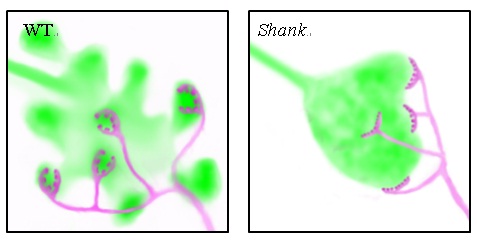
Figure1. Schematic depiction of synaptic boutons in the calyx of mushroom body at the projection neuron (green)-Kenyon cell (magenta) contact sites in wild type control (left) and Shank mutants (right). (Image by IGDB)
Contact:
Prof. ZHANG Yongqing
Institute of Genetics and Developmental Biology, Chinese Academy of Sciences